


 It’s Only Common Sense: OCCAM—the Time Is Now
 Marcy's Musings: The Growing Industry
 Dan’s Biz Bookshelf: Seeing the How
Novel Quantum Dots Enhance Cell Imaging
October 30, 2018 | University of Illinois at Urbana-ChampaignEstimated reading time: 3 minutes
A team of researchers from the University of Illinois at Urbana-Champaign and Mayo Clinic have engineered a new type of molecular probe that can measure and count RNA in cells and tissue without organic dyes. The probe is based on the conventional fluorescence in situ hybridization (FISH) technique, but it relies on compact quantum dots to illuminate molecules and diseased cells rather than fluorescent dyes.
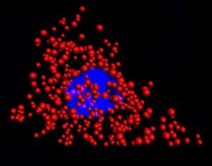
Image Caption: Quantum dots illuminate the locations of individual mRNA as red dots in the cytoplasm of a single HeLa cell. The blue region is the nucleus. This work was a collaborative effort between Illinois Bioengineering and Mayo Clinic researchers.
Over the last 50 years, FISH has evolved into a multi-billion-dollar industry because it effectively images and counts DNA and RNA in single cells. However, FISH has its limitations due to the delicate nature of the dyes. For example, the dyes rapidly deteriorate and are not very good at imaging in three dimensions. In addition, conventional FISH can only read out a couple of RNA or DNA sequences at a time.
"By replacing dyes with quantum dots, there are no stability issues whatsoever and we can count numerous RNAs with higher fidelity than before," said Andrew Smith, an associate professor of Bioengineering and member of the research team. "Moreover, we uncovered a fundamental limit to the size of a molecular label in cells, revealing new design rules for analysis in cells."
In their latest paper, published October 26, 2018, in the online edition of Nature Communications, Smith and his team identified an optimal size for quantum dots in order to effectively work with the FISH protocol. This discovery enabled quantum dot-based FISH to match the labeling accuracies currently obtained with organic dyes.
The team created unique quantum dots that are made of a zinc, selenium, cadmium, and mercury alloy and are coated with polymers. “The core of the dot dictates the wavelength of emission, and the shell dictates how much light will be given off,” said Smith, who is also affiliated with the Micro + Nanotechnology Lab, Carle Illinois College of Medicine, and Department of Materials Science and Engineering at the University of Illinois.
These dots can emit color independent of the size of the particle, which is not the case for conventional quantum dots. The dots are also small enough (7 nanometers) to fit on a probe that can maneuver between proteins and DNA in a cell, making them more comparable in size to the dyes used in conventional FISH probes.
In experiments with HeLa cells and prostate cancer cells, the researchers found that dye-based FISH cell counts declined rapidly in minutes. The quantum dot-based FISH method provided long-term luminescence to allow counting of RNA for more than 10 minutes, making it possible to acquire 3D cell imaging.
"This is important because images of cells and tissues are acquired slice-by-slice in sequence, so later slices that are labeled with dyes are depleted before they can be imaged," said Smith.
This research is part of the Mayo Illinois Alliance in which engineers from Illinois work directly with clinicians and biologists from Mayo Clinic to solve outstanding medical challenges. The Mayo Clinic Biomarker Discovery Group is working to develop FISH-based diagnostics for tumor biopsies in order to improve the accuracy of cancer diagnosis, to select personalized treatments, and to improve prognoses. The QD-FISH methodology was developed to meet this need for biomarker panels requiring the analysis of numerous genetic changes in cells in tumors for which only small amounts of specimens may be available when acquired with a fine needle, as is often the case in prostate cancer.
This research was funded by the Mayo-Illinois Alliance, National Institutes of Health, and the National Science Foundation. The full title of the paper is "Enhanced mRNA FISH with Compact Quantum Dots."
In addition to Smith, the following Illinois Bioengineering researchers contributed to the work: Yang Liu, Phuong Le, Sung Jun Lim, Liang Ma, and Suresh Sarkar. Mayo Clinic collaborators include: Dr. Farhad Kosari, Stephen Murphy, John Cheville, and George Vasmatzis.
Share on:


Suggested Items
Inkjet Solder Mask ‘Has Arrived’
04/10/2024 | Pete Starkey, I-Connect007I was delighted to be invited to attend an interactive webinar entitled “Solder Mask Coating Made Easy with Additive Manufacturing,” hosted by SUSS MicroTec Netherlands in Eindhoven. The webinar was introduced and moderated by André Bodegom, managing director at Adeon Technologies, and the speakers were Mariana Van Dam, senior product manager PCB imaging solutions at AGFA in Belgium; Ashley Steers, sales manager at Electra Polymers in the UK; and Dr. Luca Gautero, product manager at SUSS MicroTec Netherlands.
NetVia Group Acquires Direct Imaging from Mivatek
04/09/2024 | Miva TechnologiesMiva Technologies is pleased to announce NetVia Group, Irving, TX has acquired a new Miva 2400NG Dual Tray Direct Imaging System with 30-micron capabilities for inner, outer and soldermask imaging.
Teledyne to Acquire Adimec
02/13/2024 | TeledyneTeledyne Technologies Incorporated announced that it has entered into an agreement to acquire Adimec Holding B.V. and its subsidiaries.
Real Time with... productronica 2023: MivaTek Global Advances Technology With High-res Imaging System
12/08/2023 | Real Time with...productronicaMivaTek's Brendan Hogan talks about how the company employs Digitally Adaptive Rasterization Technology (DART) in their high-res imaging equipment. He also shares how the blurred line between semiconductors and microelectronics is driving broader application of the imaging process.
Keysight Enables Validation of Arbe 4D Imaging Radar Chipset
11/30/2023 | Keysight Technologies, Inc.Keysight Technologies, Inc. announces that Arbe has selected the E8719A Radar Target Solution (RTS) to test the Arbe 4D imaging radar chipset for automotive applications.