


 It’s Only Common Sense: OCCAM—the Time Is Now
 Marcy's Musings: The Growing Industry
 Dan’s Biz Bookshelf: Seeing the How
New Optical Imaging System Could Be Deployed to Find Tiny Tumors
March 8, 2019 | MITEstimated reading time: 4 minutes
Many types of cancer could be more easily treated if they were detected at an earlier stage. MIT researchers have now developed an imaging system, named “DOLPHIN,” which could enable them to find tiny tumors, as small as a couple of hundred cells, deep within the body.
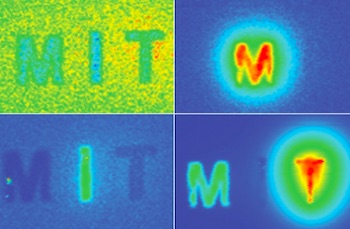
In a new study, the researchers used their imaging system, which relies on near-infrared light, to track a 0.1-millimeter fluorescent probe through the digestive tract of a living mouse. They also showed that they can detect a signal to a tissue depth of 8 centimeters, far deeper than any existing biomedical optical imaging technique.
The researchers hope to adapt their imaging technology for early diagnosis of ovarian and other cancers that are currently difficult to detect until late stages.
“We want to be able to find cancer much earlier,” says Angela Belcher, the James Mason Crafts Professor of Biological Engineering and Materials Science at MIT and a member of the Koch Institute for Integrative Cancer Research, and the newly-appointed head of MIT’s Department of Biological Engineering. “Our goal is to find tiny tumors, and do so in a noninvasive way.”
Belcher is the senior author of the study, which appears in the March 7 issue of Scientific Reports. Xiangnan Dang, a former MIT postdoc, and Neelkanth Bardhan, a Mazumdar-Shaw International Oncology Fellow, are the lead authors of the study. Other authors include research scientists Jifa Qi and Ngozi Eze, former postdoc Li Gu, postdoc Ching-Wei Lin, graduate student Swati Kataria, and Paula Hammond, the David H. Koch Professor of Engineering, head of MIT’s Department of Chemical Engineering, and a member of the Koch Institute.
Deeper Imaging
Existing methods for imaging tumors all have limitations that prevent them from being useful for early cancer diagnosis. Most have a tradeoff between resolution and depth of imaging, and none of the optical imaging techniques can image deeper than about 3 centimeters into tissue. Commonly used scans such as X-ray computed tomography (CT) and magnetic resonance imaging (MRI) can image through the whole body; however, they can’t reliably identify tumors until they reach about 1 centimeter in size.
Belcher’s lab set out to develop new optical methods for cancer imaging several years ago, when they joined the Koch Institute. They wanted to develop technology that could image very small groups of cells deep within tissue and do so without any kind of radioactive labeling.
Near-infrared light, which has wavelengths from 900 to 1700 nanometers, is well-suited to tissue imaging because light with longer wavelengths doesn’t scatter as much as when it strikes objects, which allows the light to penetrate deeper into the tissue. To take advantage of this, the researchers used an approach known as hyperspectral imaging, which enables simultaneous imaging in multiple wavelengths of light.
The researchers tested their system with a variety of near-infrared fluorescent light-emitting probes, mainly sodium yttrium fluoride nanoparticles that have rare earth elements such as erbium, holmium, or praseodymium added through a process called doping. Depending on the choice of the doping element, each of these particles emits near-infrared fluorescent light of different wavelengths.
Page 1 of 2
Share on:


Suggested Items
Lockheed Martin Successfully Transitions Long Range Discrimination Radar To The Missile Defense Agency
04/23/2024 | Lockheed MartinThe Long Range Discrimination Radar (LRDR) at Clear Space Force Station in Clear, Alaska, completed DD250 final acceptance and was officially handed over to the Missile Defense Agency in preparation for an Operational Capability Baseline (OCB) decision and final transition to the Warfighter. In addition, prior to this transition, the system has started Space Domain Awareness data collects for the United States Space Force.
US Department of Defense Selects Intel Foundry for Phase Three of RAMP-C
04/23/2024 | IntelThe U.S. Department of Defense (DoD) has awarded Intel Foundry Phase Three of its Rapid Assured Microelectronics Prototypes - Commercial (RAMP-C) program.
Real Time with... IPC APEX EXPO 2024: AI Implementation at Omron
04/18/2024 | Real Time with...IPC APEX EXPOEditor Nolan Johnson and Omron Product Manager Nick Fieldhouse discuss the company's focus on AI implementation to enhance customer experience and results. They address programming challenges and how AI can help customers achieve better outcomes with less experience. Omron's AI is compatible with existing systems, facilitating easy upgrades.
Cadence Unveils Palladium Z3 and Protium X3 Systems
04/18/2024 | Cadence Design SystemsThe Palladium Z3 and Protium X3 systems offer increased capacity, and scale from job sizes of 16 million gates up to 48 billion gates, so the largest SoCs can be tested as a whole rather than just partial models, ensuring proper functionality and performance.
Real Time with... IPC APEX EXPO 2024: MYCRONIC's Evolution and New Solutions
04/17/2024 | Real Time with...IPC APEX EXPOHenry Crandall interviews Kevin Clue, the vice president of global sales for MYCRONIC's High Flex division. They discuss the company's evolution, emphasizing its strong customer relationships and its role as a versatile, turnkey solution provider. Kevin unveils new solutions launched at IPC APEX EXPO, including an AI-integrated inspection system and the A40 pick-and-place platform. The conversation also touches on the increased use of AI and deep learning.