


 It’s Only Common Sense: OCCAM—the Time Is Now
 Marcy's Musings: The Growing Industry
 Dan’s Biz Bookshelf: Seeing the How
Pen-sized Microscope could ID Cancer Cells in Doctor's Offices and Operating Rooms
January 27, 2016 | University of WashingtonEstimated reading time: 3 minutes
Surgeons removing a malignant brain tumor don’t want to leave cancerous material behind. But they’re also trying to protect healthy brain matter and minimize neurological harm.
Once they open up a patient’s skull, there’s no time to send tissue samples to a pathology lab — where they are typically frozen, sliced, stained, mounted on slides and investigated under a bulky microscope — to definitively distinguish between cancerous and normal brain cells.
But a handheld, miniature microscope being developed by University of Washington mechanical engineers could allow surgeons to “see” at a cellular level in the operating room and determine where to stop cutting.
The new technology, developed in collaboration with Memorial Sloan Kettering Cancer Center, Stanford University and the Barrow Neurological Institute, is outlined in a paper published in January in the journal Biomedical Optics Express.
“Surgeons don’t have a very good way of knowing when they’re done cutting out a tumor,” said senior author Jonathan Liu, UW assistant professor of mechanical engineering. “They’re using their sense of sight, their sense of touch, pre-operative images of the brain — and oftentimes it’s pretty subjective.
“Being able to zoom and see at the cellular level during the surgery would really help them to accurately differentiate between tumor and normal tissues and improve patient outcomes,” said Liu.
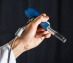
The handheld microscope, roughly the size of a pen, combines technologies in a novel way to deliver high-quality images at faster speeds than existing devices. Researchers expect to begin testing it as a cancer-screening tool in clinical settings next year.
UW mechanical engineering doctoral students and assistant professor Jonathan T.C. Liu work to align a handheld microscope for cancer detection in patients. From left to right: Ye Chen, Linpeng “Peter” Wei, Liu and Chengbo Yin.Dennis Wise, University of Washington
For instance, dentists who find a suspicious-looking lesion in a patient’s mouth often wind up cutting it out and sending it to a lab to be biopsied for oral cancer. Most come back benign.
That process subjects patients to an invasive procedure and overburdens pathology labs. A miniature microscope with high enough resolution to detect changes at a cellular level could be used in dental or dermatological clinics to better assess which lesions or moles are normal and which ones need to be biopsied.
To create a handheld dual-axis confocal microscope, UW engineers miniaturized the larger microscope prototype seen on the table into a device roughly the size of a pen.Dennis Wise, University of Washington
“The microscope technologies that have been developed over the last couple of decades are expensive and still pretty large, about the size of a hair dryer or a small dental x-ray machine,” said co-author Milind Rajadhyaksha, associate faculty member in the dermatology service at the Memorial Sloan Kettering Cancer Center in New York City. “So there’s a need for creating much more miniaturized microscopes.”
Making microscopes smaller, however, usually requires sacrificing some aspect of image quality or performance such as resolution, field of view, depth, imaging contrast or processing speed.
“We feel like this device does one of the best jobs ever — compared to existing commercial devices and previous research devices — of balancing all those tradeoffs,” said Liu.
The miniature microscope uses an innovative approach called “dual-axis confocal microscopy” to illuminate and more clearly see through opaque tissue. It can capture details up to a half millimeter beneath the tissue surface, where some types of cancerous cells originate.
In the video below, for instance, researchers produced images of fluorescent blood vessels in a mouse ear at various depths ranging from 0.075 to 0.125 millimeters deep.
Share on:


Suggested Items
Inkjet Solder Mask ‘Has Arrived’
04/10/2024 | Pete Starkey, I-Connect007I was delighted to be invited to attend an interactive webinar entitled “Solder Mask Coating Made Easy with Additive Manufacturing,” hosted by SUSS MicroTec Netherlands in Eindhoven. The webinar was introduced and moderated by André Bodegom, managing director at Adeon Technologies, and the speakers were Mariana Van Dam, senior product manager PCB imaging solutions at AGFA in Belgium; Ashley Steers, sales manager at Electra Polymers in the UK; and Dr. Luca Gautero, product manager at SUSS MicroTec Netherlands.
NetVia Group Acquires Direct Imaging from Mivatek
04/09/2024 | Miva TechnologiesMiva Technologies is pleased to announce NetVia Group, Irving, TX has acquired a new Miva 2400NG Dual Tray Direct Imaging System with 30-micron capabilities for inner, outer and soldermask imaging.
Teledyne to Acquire Adimec
02/13/2024 | TeledyneTeledyne Technologies Incorporated announced that it has entered into an agreement to acquire Adimec Holding B.V. and its subsidiaries.
Real Time with... productronica 2023: MivaTek Global Advances Technology With High-res Imaging System
12/08/2023 | Real Time with...productronicaMivaTek's Brendan Hogan talks about how the company employs Digitally Adaptive Rasterization Technology (DART) in their high-res imaging equipment. He also shares how the blurred line between semiconductors and microelectronics is driving broader application of the imaging process.
Keysight Enables Validation of Arbe 4D Imaging Radar Chipset
11/30/2023 | Keysight Technologies, Inc.Keysight Technologies, Inc. announces that Arbe has selected the E8719A Radar Target Solution (RTS) to test the Arbe 4D imaging radar chipset for automotive applications.